Human Skin Cell Under Microscope Micropedia Images and Photos finder
Mitosis in an animal cell. Cells from the Chinese Hamster Ovary are shown undergoing mitosis. Beginning with a cell spread on the substrate, follow prophase,.

Cheek Cell Lab Aiden's Blog
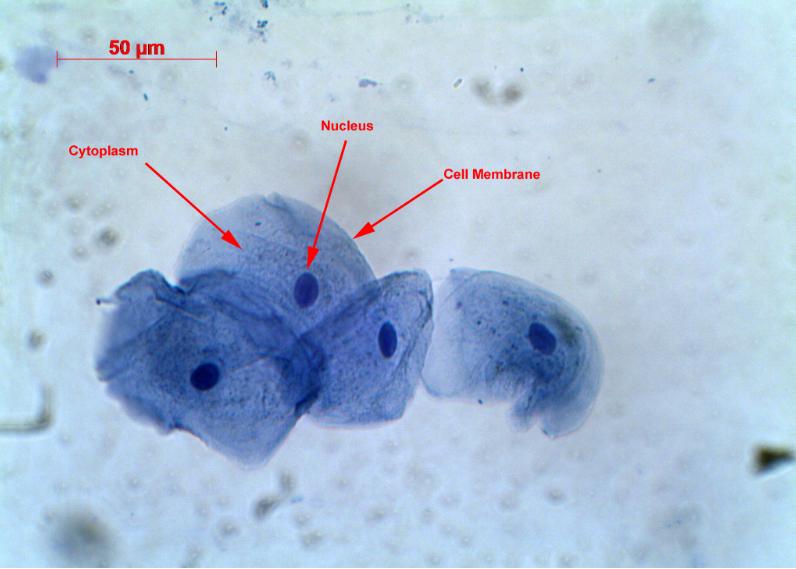
Human cheek cells are made of simple squamous epithelial cells, which are flat cells with a round visible nucleus that cover the inside lining of the cheek.C.
Some cells under the microscope. 大鲵小站
A typical animal cell is 10-20 μm in diameter, which is about one-fifth the size of the smallest particle visible to the naked eye. It was not until good light microscopes became available in the early part of the nineteenth century that all plant and animal tissues were discovered to be aggregates of individual cells. This discovery, proposed as the cell doctrine by Schleiden and Schwann.
Are we really made up of microscopic cells? conspiracy
How does the human body look like under an electron microscope?Objects Under Electron Microscope! (Part - 1). Watch Here https://youtu.be/c6Jqis6wrbkMusic: h.

And these are our lung cells. Microscopic photography, Things under a microscope, Biology art
Remove any excess solution by allowing a paper towel to touch one side of the coverslip. Place the slide on the microscope, with 4 x or 10 x objective in position and find a cell. Then view at higher magnification. Methylene blue stains negatively charged molecules in the cell, including DNA and RNA. This dye is toxic when ingested and it.
4.2 Discovery of Cells and Cell Theory Human Biology
Cells and Tissues. Tissues are classified into four basic types: epithelium, connective tissue (includes cartilage, bone and blood), muscle, and nervous tissue. Organs are assembled from the four basic types of tissues and have cells with specialized functions. These slides contain tissue sections that are easily confused with each other.

Scientists developed a microscope that fits in a needle to get a realtime look inside the human
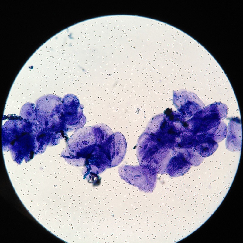
Cheek Cells Under a Microscope Requirements, Preparation and Staining Cheek cells are eukaryotic cells (cells that contain a nucleus and other organelles within enclosed in a membrane) that are easily shed from the mouth lining. It's therefore easy to obtain them for observation. Some of the main parts of a cell include: 1.

Human Cells Under Microscope HighRes Stock Photo Getty Images
Microscopy Introduction to microscopes and how they work. Covers brightfield microscopy, fluorescence microscopy, and electron microscopy. Introduction If you meet some cell biologists and get them talking about what they enjoy most in their work, you may find it comes down to one thing: secretly, they're all microscope freaks.
Cells Rumney Marsh Academy Science Revere, Massachusetts
In Figure 3.1.2 3.1. 2, only one edge of the tissue slice has epithelial cells. In Figure 3.1.2 3.1. 2 A that edge is indicated with an arrow, but when looking at a specimen under a microscope, you have to figure out for yourself where the edge with the epithelial cells is. Figure 3.1.2 3.1. 2: A slice of a trachea.
February 2011 Cell As a Unit of Life
Browse 1,185 authentic human cells under microscope stock videos, stock footage, and video clips available in a variety of formats and sizes to fit your needs, or explore cancer cells or dna stock videos to discover the perfect clip for your project. Browse Getty Images' premium collection of high-quality, authentic Human Cells Under Microscope.

10,151 Human Cell Under Microscope Images, Stock Photos & Vectors Shutterstock
The Human Cheek Cell. 1. List the 3 parts of the Cell Theory. 2. Describe or define each of the following. Draw your cells to scale. 4. Why is methylene blue necessary? 5. The light microscope used in the lab is not powerful enough to view other organelles in the cheek cell.. We also acknowledge previous National Science Foundation.
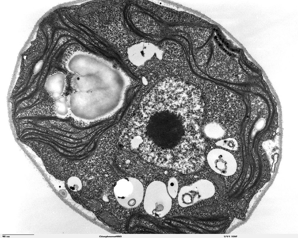
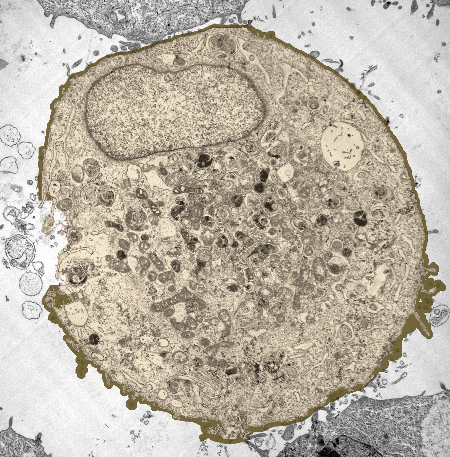
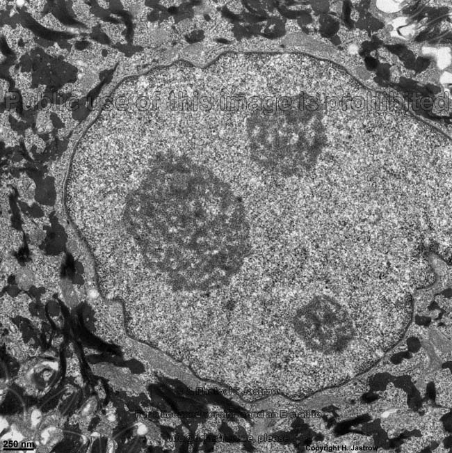
Electron Microscopy of a normal human cell, The cell membrane, nucleus and the nucleolus are all
The Human Body Under the Microscope | Discover Magazine The Sciences Mind Technology Health Environment Planet Earth Lifestyle / Planet Earth The Human Body Under the Microscope A visual voyage through the cells, organs, microbes and molecules that make up our bodies. By Colin Salter Feb 11, 2015 10:00 AMNov 18, 2019 4:30 PM Newsletter

Researchers Identify Protein that Makes Skin Cancer Cells More Invasive Onco'Zine
Human Cheek Cells Figure 3. Human cheek cell at 400x zoom.. View under the microscope using the highest magnification for the best cellular details and draw what you see. Be sure to indicate the magnification used and specimen name. Also, indicate the estimated cell size in micrometers under your drawing. Figure 7.
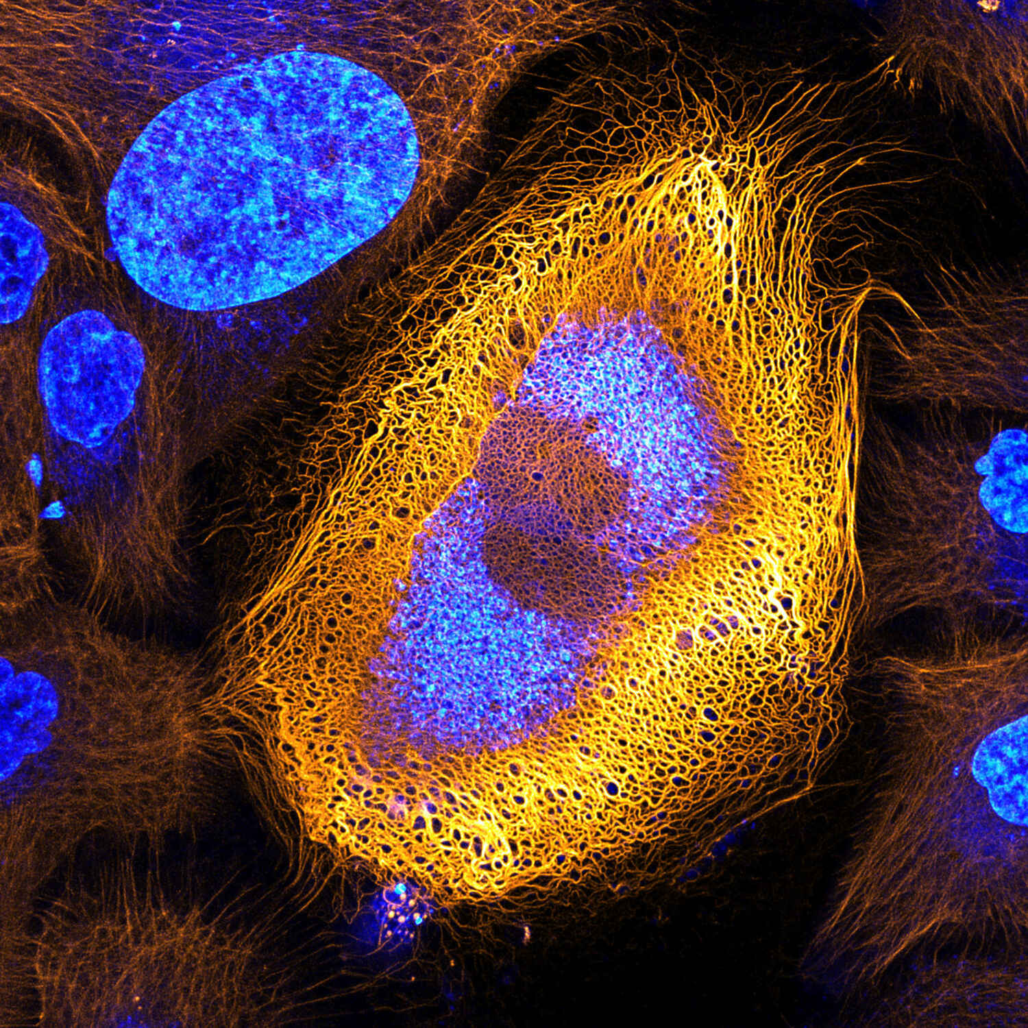
Stunning Microscopic View of Human Skin Cells Wins 2017 Nikon Small World Competition News
Anton van Leeuwenhoek was the first person to observe living cells under the microscope in 1675—he described many types of cells, including bacteria. Since then more sophisticated and powerful scopes have been developed that allow for higher magnification and clearer images.. This includes human cells and many other types of cells that you.

blood cells, cells, human, electron microscope, scan, blood, microscopic, medicine, microbiology
Observing human cheek cells under a microscope is a simple way to quickly view and learn about human cell structure.
Cell Under Electron Microscope Video Bokep Ngentot
Browse 2,752 human cell under microscope photos and images available, or start a new search to explore more photos and images. of 46 NEXT Browse Getty Images' premium collection of high-quality, authentic Human Cell Under Microscope stock photos, royalty-free images, and pictures.